Home

avion izolare pernă proteine microscop Nefavorabil piersică Sens tactil

Protein Analysis Techniques Explained - ATA Scientific

Electron micrographs of the spike protein of Sars-CoV-2 - YouTube

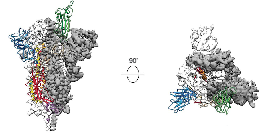
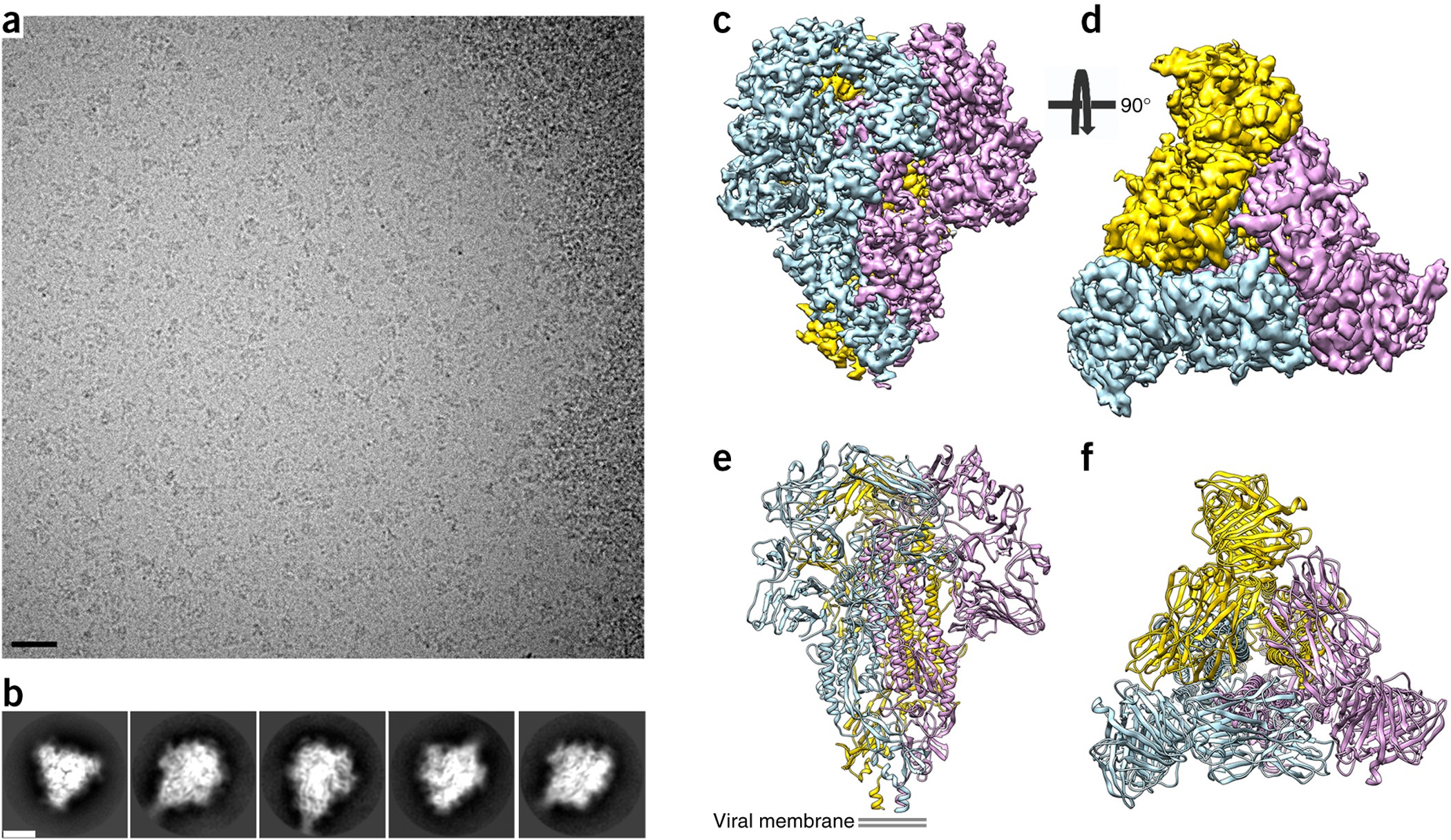
Glycan shield and epitope masking of a coronavirus spike protein observed  by cryo-electron microscopy | Nature Structural & Molecular Biology
Glycan shield and epitope masking of a coronavirus spike protein observed by cryo-electron microscopy | Nature Structural & Molecular Biology

Electron microscopy images of protein structures in purified fractions... |  Download Scientific Diagram
Electron microscopy images of protein structures in purified fractions... | Download Scientific Diagram
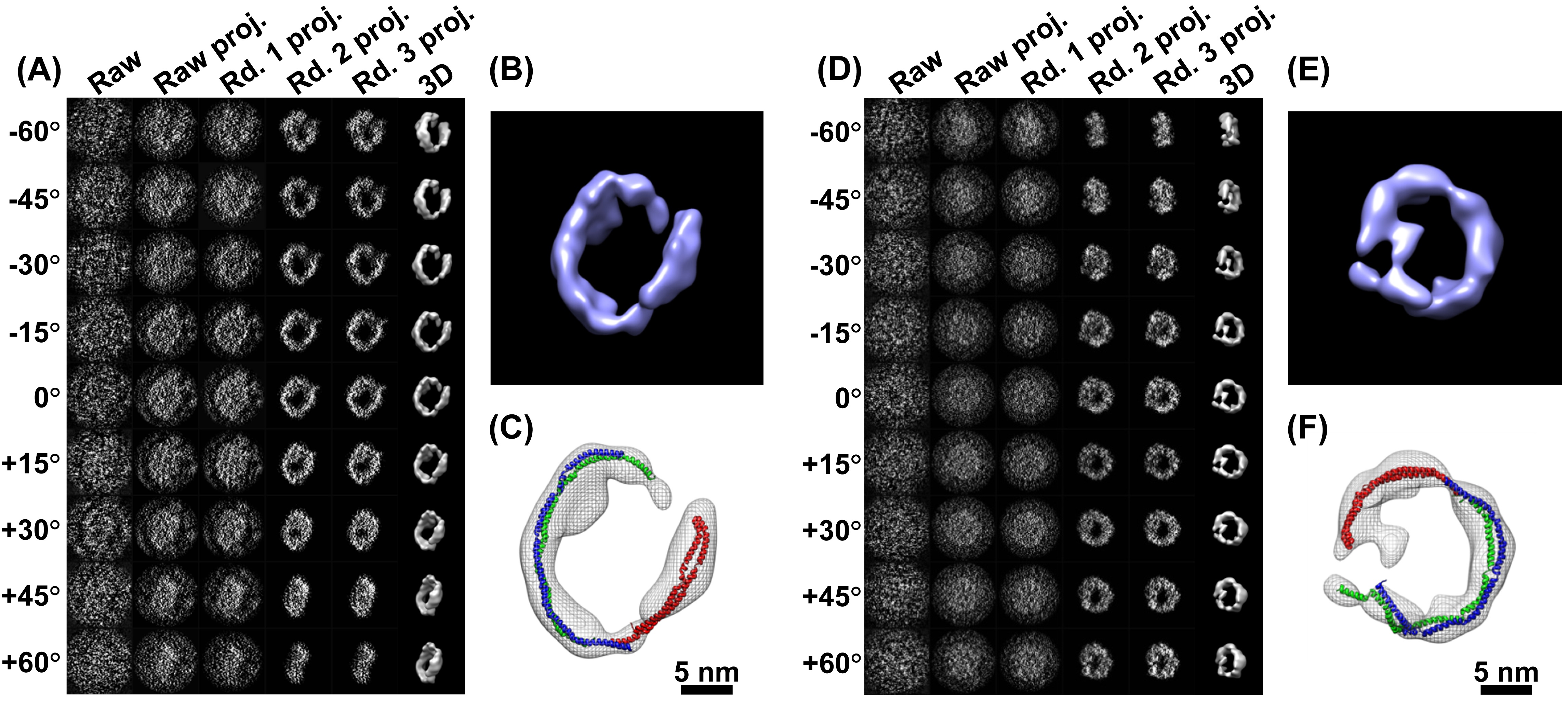
Referat Chimie PROTEINELE | PDF

Under the Electron Microscope – A 3-D Image of an Individual Protein -  Berkeley Lab – Berkeley Lab News Center
Under the Electron Microscope – A 3-D Image of an Individual Protein - Berkeley Lab – Berkeley Lab News Center

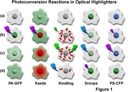
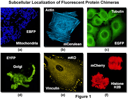
Confocal Microscopy - Optical Highlighter Fluorescent Proteins | Olympus LS

PDB-101: Learn: Guide to Understanding PDB Data: Methods for Determining  Structure
PDB-101: Learn: Guide to Understanding PDB Data: Methods for Determining Structure

Microscopie fluorescentă - Wikipedia

Super-resolution microscopy to visualize and quantify protein  microstructural organization in food materials and its relation to  rheology: Egg white proteins - ScienceDirect
Super-resolution microscopy to visualize and quantify protein microstructural organization in food materials and its relation to rheology: Egg white proteins - ScienceDirect

Comment observe-t-on les protéines à l'atome près ?

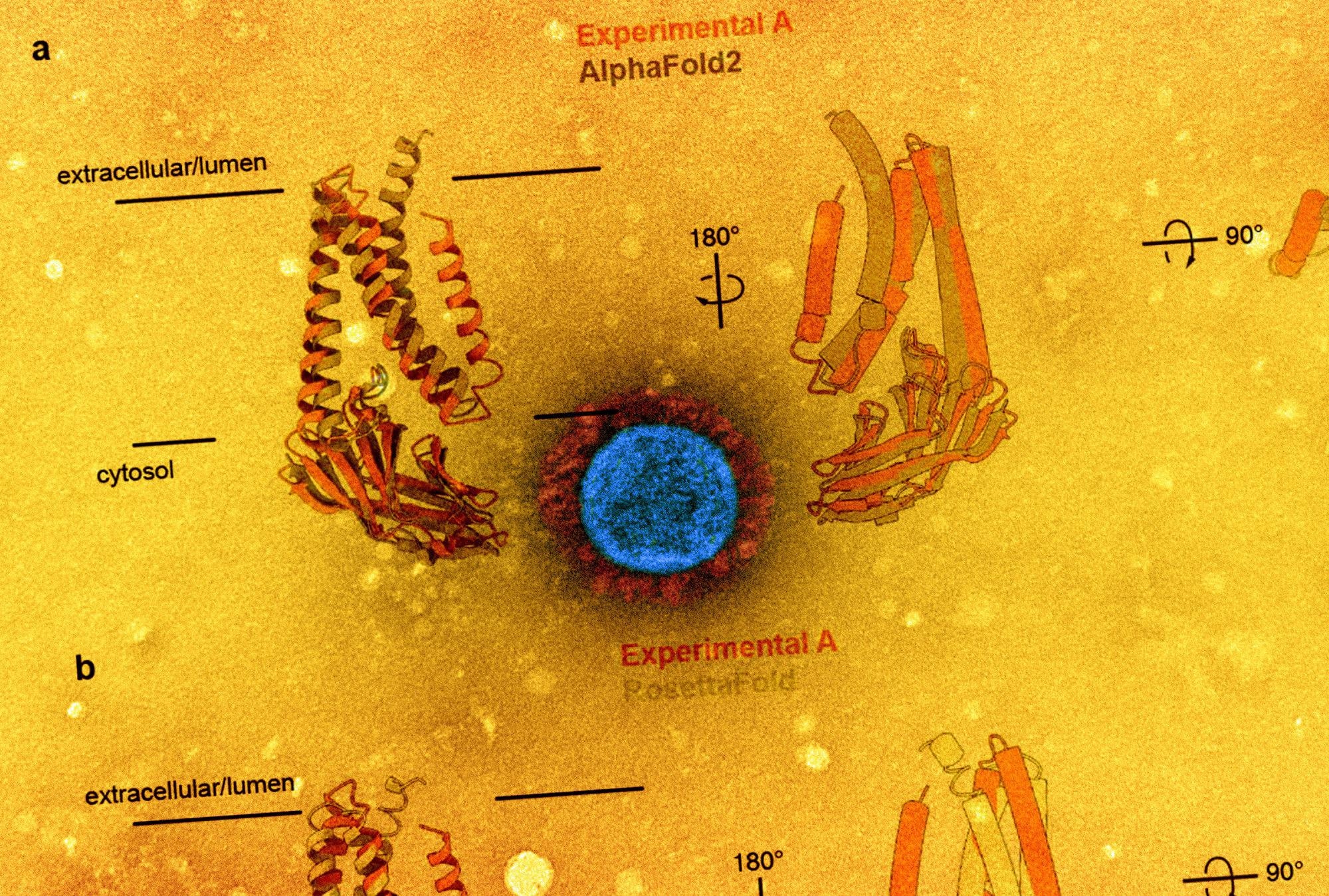
The Coronavirus Unveiled: Microscopic Images of SARS-CoV-2 - The New York  Times
The Coronavirus Unveiled: Microscopic Images of SARS-CoV-2 - The New York Times

A new glow for electron microscopy

First glimpses of motor proteins in action | Corporate | University of Leeds

Proteinele | Sanatate si nutritie

Scanning electron microscopy image of outer membrane protein (OMP) of... |  Download Scientific Diagram
Scanning electron microscopy image of outer membrane protein (OMP) of... | Download Scientific Diagram
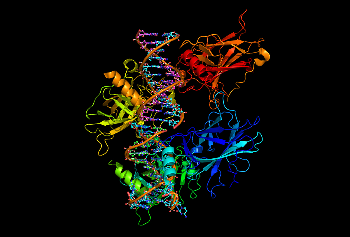
First glimpses of motor proteins in action | Corporate | University of Leeds

Nanomasinile umane
Nanomasinile umane

Cancere tratate cu proteine artificiale. Cercetătorii anunţă o nouă terapie  | adevarul.ro
Cancere tratate cu proteine artificiale. Cercetătorii anunţă o nouă terapie | adevarul.ro

Light microscopy provides a deep look into protein structure | FAU  Erlangen-Nürnberg
Light microscopy provides a deep look into protein structure | FAU Erlangen-Nürnberg

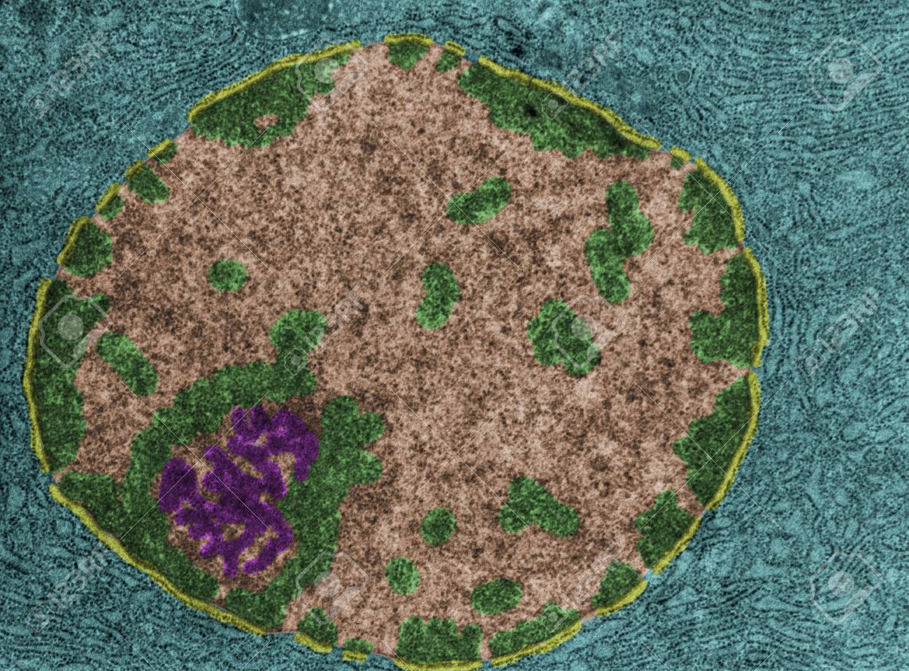
False Colour Transmission Electron Microscope (TEM) Showing The Nucleus Of  A Protein-synthesizing Cell. The Nuclear Envelope (yellow), Chromatin  (green), Nucleoplasm (light Brown) And Nucleolus (magenta) Can Be Seen. The  Cytoplasm (cian) Is
False Colour Transmission Electron Microscope (TEM) Showing The Nucleus Of A Protein-synthesizing Cell. The Nuclear Envelope (yellow), Chromatin (green), Nucleoplasm (light Brown) And Nucleolus (magenta) Can Be Seen. The Cytoplasm (cian) Is

Supercool Protein Imaging Gets the Nobel Prize | Quanta Magazine

Cryo-electron microscopy reveals structures of protein that maintains cell  membranes
Cryo-electron microscopy reveals structures of protein that maintains cell membranes

Cryo-electron microscopy structure of SARS-CoV-2 membrane protein

Confocal Microscopy - The Fluorescent Protein Color Palette | Olympus LS

The SARS-CoV spike protein and its receptor - Healthcare industry